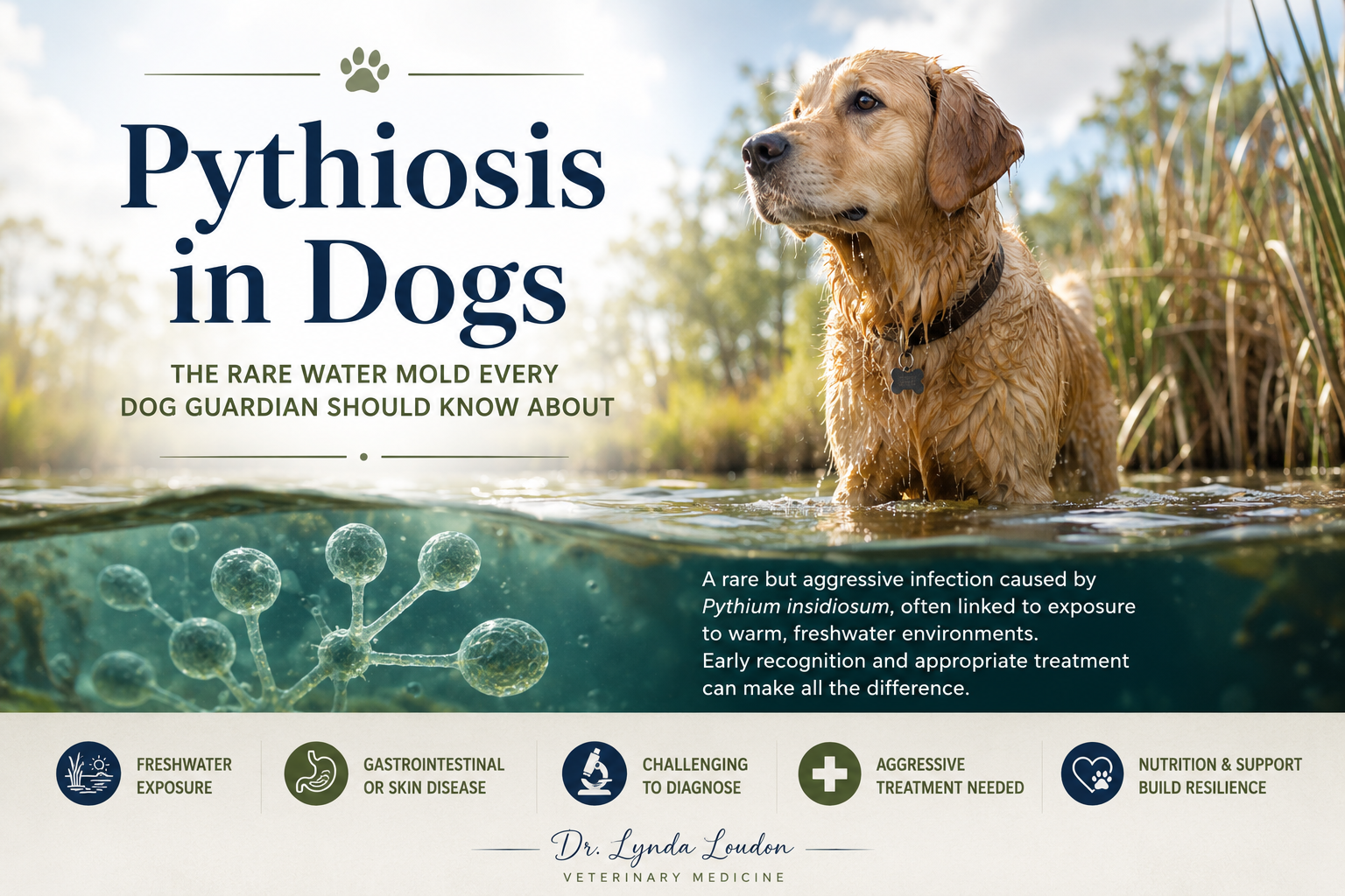
When most dog guardians think about infections, they think of bacteria, viruses, or fungi. But there is another organism that, while rare, can cause devastating disease in dogs: Pythium insidiosum.
Pythiosis is one of those diseases that often isn’t recognized until a dog has been sick for weeks or even months. Because it can mimic inflammatory bowel disease, cancer, or chronic skin infections, it frequently flies under the radar.
The good news? Early recognition can make a tremendous difference.
What is Pythiosis?
Pythiosis is caused by Pythium insidiosum, an organism often referred to as a “water mold.” Although it’s commonly mistaken for a fungus, it actually belongs to a completely different group of organisms called oomycetes, which are more closely related to algae than fungi.
This distinction is important because many antifungal medications that work well against true fungi are much less effective against Pythium.
How Do Dogs Become Infected?
Dogs typically become infected after exposure to warm, stagnant freshwater.
Common sources include:
- Ponds
- Marshes
- Swamps
- Flooded fields
- Slow-moving streams
- Irrigation ditches
The organism produces swimming spores that enter the body through small wounds in the skin or are swallowed while a dog is drinking or swimming.
Dogs living in warm, humid climates are at greatest risk, but cases have been reported in many regions.
Two Forms of Disease
Gastrointestinal Pythiosis
This is the most common form in dogs.
Signs may include:
- Chronic vomiting
- Chronic diarrhea
- Weight loss
- Poor appetite
- Abdominal pain
- Thickened intestines
- Intestinal masses
Unfortunately, these signs are very similar to inflammatory bowel disease, lymphoma, or other chronic gastrointestinal disorders.
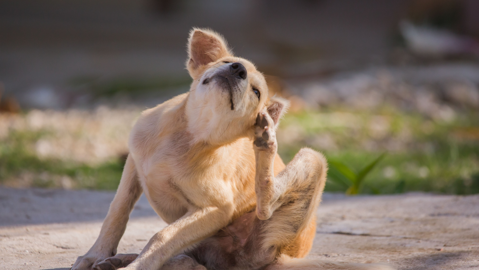
Cutaneous (Skin) Pythiosis
Some dogs develop skin infections instead.
These dogs may have:
- Draining wounds
- Firm nodules
- Ulcers
- Swelling that won’t heal
- Chronic draining tracts
How Is Pythiosis Diagnosed?
One of the biggest challenges with pythiosis is that there isn’t a single blood test or imaging study that can diagnose it on its own. Instead, veterinarians piece together the diagnosis using your dog’s history, physical examination, imaging findings, tissue biopsies, and specialized laboratory testing.
Step 1: Recognizing the Possibility
The first step is simply thinking about pythiosis as a possible diagnosis.
If your dog has been exposed to warm, stagnant freshwater such as ponds, marshes, swamps, flooded fields, or slow-moving streams and develops chronic gastrointestinal disease or a wound that refuses to heal, pythiosis should be on the list of possible causes.
Dogs with the gastrointestinal form often develop:
- Chronic vomiting
- Chronic diarrhea
- Weight loss
- Decreased appetite
- Thickening of the intestines or an intestinal mass
Dogs with the skin form may develop:
- Draining wounds
- Firm nodules
- Ulcerated skin lesions
- Areas that fail to heal despite antibiotics
Because these signs can mimic inflammatory bowel disease, fungal infections, cancer, or chronic bacterial infections, a high level of suspicion is often what leads to the correct diagnosis.
Step 2: Imaging
Abdominal ultrasound is usually one of the first diagnostic tools used when gastrointestinal pythiosis is suspected.
Your veterinarian may identify:
- Thickened intestinal walls
- Loss of the normal intestinal layers
- Intestinal masses
- Enlarged lymph nodes
- Partial intestinal obstruction
These findings are not specific for pythiosis, but they help determine where the disease is located and guide biopsy collection.
If surgery is being considered, chest X-rays or a CT scan may also be recommended to help assess the extent of disease and assist with surgical planning.
Step 3: Bloodwork
Routine bloodwork helps evaluate your dog’s overall health but cannot diagnose pythiosis.
Depending on how advanced the disease is, bloodwork may reveal:
- Mild anemia
- Increased eosinophils (a type of white blood cell often associated with parasites or certain inflammatory diseases)
- Low albumin due to protein loss from the intestines
Many dogs have fairly nonspecific laboratory changes, which is one reason this disease can be challenging to recognize early.
Step 4: Tissue Biopsy — The Most Important Step
A tissue biopsy is considered the cornerstone of diagnosis.
For dogs with intestinal disease, a full-thickness surgical biopsy is often preferred because the organism tends to invade the deeper layers of the intestinal wall. Small endoscopic biopsies may miss the disease altogether.
For dogs with skin lesions, a deep biopsy taken from the active edge of the lesion provides the best chance of making a diagnosis.
A veterinary pathologist examines the tissue under the microscope, looking for:
- Granulomatous or pyogranulomatous inflammation
- Large numbers of eosinophils
- Areas of tissue destruction
- Broad, sparsely septate hyphae surrounded by a characteristic inflammatory reaction known as Splendore-Hoeppli material
Special stains, such as Gomori methenamine silver (GMS) or Periodic acid-Schiff (PAS), help make the organism much easier to identify.
Step 5: Antibody Testing (Serology)
One of the most helpful diagnostic tools is a Pythium antibody test (ELISA).
This blood test looks for antibodies your dog’s immune system has produced against Pythium insidiosum.
When interpreted alongside the dog’s history, clinical signs, and biopsy results, this test has excellent sensitivity and specificity.
It can also be useful for monitoring treatment, as antibody levels often decrease when treatment is successful and may rise again if the disease returns.
Step 6: PCR and Culture
In some cases, additional testing may be performed.
Specialized laboratories can use PCR testing to detect Pythium DNA within biopsy tissue, providing further confirmation of the diagnosis.
The organism can also be cultured from tissue samples, although this is technically difficult, not always successful, and is generally reserved for specialty or research laboratories.
Putting It All Together
No single test confirms pythiosis in every patient. Instead, veterinarians combine your dog’s history, imaging findings, biopsy results, and specialized laboratory testing to arrive at the diagnosis.
The most important message for dog guardians is:
If your dog has chronic gastrointestinal disease or a wound that simply won’t heal and they’ve spent time in warm freshwater ask your veterinarian whether pythiosis should be considered.
Because this disease can closely resemble inflammatory bowel disease, fungal infections, or even cancer, recognizing it early can significantly improve your dog’s chances of successful treatment.
Why Is Pythiosis So Difficult to Treat?
Unlike true fungi, Pythium doesn’t have the same cell membrane that many antifungal drugs target.
As a result, medications that are effective for fungal infections often provide only limited benefit against pythiosis.
The best outcomes occur when the disease is diagnosed early enough for complete surgical removal of the affected tissue.
Treatment may include:
- Surgery
- Antifungal medications such as terbinafine and itraconazole
- Pythium immunotherapy (available in some cases)
- Supportive care
Can Diet Help?
This is a question I hear often.
Unfortunately, there is no diet that has been shown to eliminate Pythium.
However, nutrition plays an incredibly important role in supporting the dog’s overall resilience during treatment.
My priorities would be:
- Feed a balanced, fresh, highly digestible whole food diet.
- Maintain lean muscle mass.
- Support the gut microbiome with appropriate probiotics and prebiotic fibers.
- Provide omega-3 fatty acids to help regulate inflammation.
- Consider medicinal mushrooms, such as turkey tail or reishi, as immune-supportive therapies.
- Individualize the diet based on the dog’s digestive tolerance.
For dogs with significant intestinal disease, a gently cooked fresh diet is often easier to digest than raw. While I often recommend raw diets, they are not appropriate for every dog.
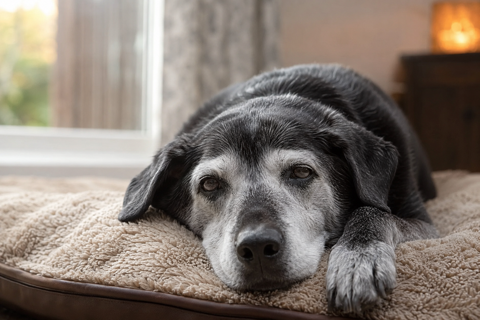
Building Resilience During Recovery
One of the principles I teach is that resilience matters most when the body is under stress.
A dog recovering from pythiosis benefits from support that extends beyond medications alone.
That includes:
- Excellent nutrition
- Maintaining hydration
- Protecting the gut microbiome
- Controlling inflammation
- Minimizing unnecessary medications when appropriate
- Supporting emotional well-being and reducing stress
These measures won’t cure pythiosis, but they help support the body’s ability to heal.
Prognosis
The prognosis depends largely on:
- How early the disease is diagnosed
- Whether complete surgical removal is possible
- Whether the infection is localized or widespread
Dogs whose lesions can be completely removed surgically generally have a much better outlook than those with extensive intestinal involvement.
My Take
Pythiosis reminds us why it’s so important not to settle for “it’s probably just a sensitive stomach” when a dog has persistent gastrointestinal signs.
If your dog has chronic vomiting, diarrhea, unexplained weight loss, or an intestinal mass, especially if they have been exposed to warm freshwater, ask your veterinarian whether pythiosis should be on the list of possible diagnoses.
It’s rare, but it’s one of those diseases where recognizing it early can make all the difference.
Remember: Every decision you make either builds your dog’s resilience—or slowly erodes it. While no supplement or diet can cure pythiosis, providing exceptional nutrition, supporting the microbiome, and partnering with an experienced integrative veterinarian can help give your dog the best possible chance at recovery.